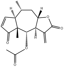
生物活性
Bigelovin 是可从海百合中分离得到的一种倍半萜内酯,是选择性的视黄素 X 受体 α (retinoid X receptor α) 的激动剂。Bigelovin 可通过诱导凋亡和自噬来发挥抗肿瘤活性。Bigelovin 通过抑制 ROS 的生成来调节 mTOR 信号通路。
体外研究
Bigelovin (0-20 μM, 24-72 h) significantly inhibits cell viability of liver cancer cells and induces apoptosis and autophagy.
Bigelovin causes a significant increase of p62, LC3B-II, Beclin-1 and a corresponding decrease of p62 levels in a time-dependent manner.
Bigelovin induces cell death involves the suppression of mTOR pathway regulated by ROS production.
Cell Viability Assay
Cell Line: HepG2 and SMMC-7721 cells.
Concentration: 0-20 μM.
Incubation Time: 24, 48, 72 h.
Result: Significantly reduced the cell viability of HepG2 and SMMC-7721 cells in a dose- and time dependent manner.
No significant difference observed in cell viability of normal liver cell lines, LO2 and LX2, after BigV treatment for 24, 48 or 72 h.
Western Blot Analysis
Cell Line: HepG2 and SMMC-7721 cells.
Concentration: 0-10 μM.
Incubation Time: 24 h.
Result: The expression of Bcl-2 was decreased, whereas Bax was increased after treatment with BigV. Moreover, Caspase-9, -3 and PARP cleavage were activated significantly after BigV treatment.
Cell Line:
HepG2 and SMMC-7721 cells.
Incubation Time:
24, 48, 72 h.
Result:
Significantly reduced the cell viability of HepG2 and SMMC-7721 cells in a dose- and time dependent manner.
No significant difference observed in cell viability of normal liver cell lines, LO2 and LX2, after BigV treatment for 24, 48 or 72 h.
Cell Line:
HepG2 and SMMC-7721 cells.
Result:
The expression of Bcl-2 was decreased, whereas Bax was increased after treatment with BigV. Moreover, Caspase-9, -3 and PARP cleavage were activated significantly after BigV treatment.
体内研究
Bigelovin (BigV, 5, 10, 20 mg/kg) exerts anti-tumor activity in HepG2 xenograft tumor model.
Animal Model: HepG2 xenograft model based on the male athymic BALB/c nude mice (5-6 weeks old, 18-22 g).
Dosage: 5, 10, 20 mg/kg.
Administration: Intravenous injection every 2 days.
Result: The tumor growth rate was significantly slower in BigV treated groups in a dose-dependent manner, along with the reduced tumor weight.
No significant alteration of body weight and hepatic enzyme levels (AST, ALT and LDH) in serum was observed after BigV administration.
Western blot findings of tumor tissues indicated the activation of apoptosis and autophagy characterized by the increase of cleaved Caspase-3 and PARP, as well as LC3BII levels.
The inactivation of mTOR was also observed in tumor tissues isolated from BigV-treated mice.
Animal Model:
HepG2 xenograft model based on the male athymic BALB/c nude mice (5-6 weeks old, 18-22 g).
Administration:
Intravenous injection every 2 days.
Result:
The tumor growth rate was significantly slower in BigV treated groups in a dose-dependent manner, along with the reduced tumor weight.
No significant alteration of body weight and hepatic enzyme levels (AST, ALT and LDH) in serum was observed after BigV administration.
Western blot findings of tumor tissues indicated the activation of apoptosis and autophagy characterized by the increase of cleaved Caspase-3 and PARP, as well as LC3BII levels.
The inactivation of mTOR was also observed in tumor tissues isolated from BigV-treated mice.

 15623309010


