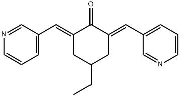
4-乙基-2,6-双(吡啶-3-基亚甲基)环己酮
中文名称:
4-乙基-2,6-双(吡啶-3-基亚甲基)环己酮
中文同义词:
(2E,6E)-4-乙基-2,6-二(3-吡啶基亚甲基)环己酮;4-乙基-2,6-双(吡啶-3-基亚甲基)环己酮;MCB-613游离态
英文名称:
MCB-613
英文同义词:
4-ethyl-2,6-bis(pyridin-3-ylmethylene)cyclohexanone;(2E,6E)-4-Ethyl-2,6-bis(3-pyridinylmethylene)cyclohexanone;MCB-613;CS-2522;(2E,6E)-4-ethyl-2,6-bis(pyridin-3-ylmethylidene)cyclohexan-1-one;Cyclohexanone, 4-ethyl-2,6-bis(3-pyridinylmethylene)-, (2E,6E)-
CAS号:
1162656-22-5
分子式:
C20H20N2O
分子量:
304.39
EINECS号:
相关类别:
合成材料中间体;API
Mol文件:
1162656-22-5.mol
沸点
510.4±50.0 °C(Predicted)
密度
1.159±0.06 g/cm3(Predicted)
储存条件
Sealed in dry,Room Temperature
酸度系数(pKa)
4.36±0.12(Predicted)
生物活性
MCB-613 是一种有效的类固醇受体共激活因子 (SRC) 小分子“刺激物” (SMS),超刺激 SRCs 的转录活性。 MCB-613 增加了 SRC 与其他共激活因子的相互作用,并显着诱导 ER 应激与活性氧 (ROS) 的产生相结合。 MCB-613 是一种 SMS,通过过度刺激 SRC 致癌程序,靶向癌基因用作抗癌药物。
体外研究
MCB-613 (6-8 μM; 24 hours) activates endogenous MMP13 mRNA expression in MDA-MB-231 cells. MCB-613 (2-10 μM; 4 hours) leads to proteasome dysfunction and ER stress, the induction of the markers for unfolded protein response (UPR), including the phosphorylation of eIF2α and IRE1α as well as the induction of ATF4 protein expression. MCB-613 (0-7 μM; 4 hours) affects SRC-3 KO and WT HeLa cell viability, SRC-3 WT HeLa cell is more affected by MCB-613 compared with KO cells.
RT-PCR
Cell Line: MDA-MB-231 cells
Concentration: 6 μM; 8 μM
Incubation Time: 24 hours
Result: Increased MMP13 mRNA expression.
Western Blot Analysis
Cell Line: HeLa cells
Concentration: 2 μM; 4 μM; 6 μM; 8 μM; 10 μM
Incubation Time: 24 hours
Result: Induced the p-eIF2α, p-IRE1α, and ATF-4 protein expression.
Cell Viability Assay
Cell Line: SRC-3 KO and WT HeLa cells
Concentration: 3 μM; 4 μM; 5 μM; 6 μM; 7 μM
Incubation Time: 24 hours
Result: Decreased SRC-3 KO and WT HeLa cell viability.
Cell Line:
MDA-MB-231 cells
Concentration:
6 μM; 8 μM
Incubation Time:
24 hours
Result:
Increased MMP13 mRNA expression.
Cell Line:
HeLa cells
Concentration:
2 μM; 4 μM; 6 μM; 8 μM; 10 μM
Incubation Time:
24 hours
Result:
Induced the p-eIF2α, p-IRE1α, and ATF-4 protein expression.
Cell Line:
SRC-3 KO and WT HeLa cells
Concentration:
3 μM; 4 μM; 5 μM; 6 μM; 7 μM
Incubation Time:
24 hours
Result:
Decreased SRC-3 KO and WT HeLa cell viability.
体内研究
MCB-613 (intravenous injection; 20 mg/kg; 3 times/week; 7 weeks) significantly and dramatically stalls the growth of the tumor compared with the control group and causes no obvious animal toxicity
Animal Model: MCF-7 breast cancer mouse xenograft model (athymic nude mice by injecting MCF-7 cells into mammary fat pads)
Dosage: 20 mg/kg
Administration: Intravenous injection; 20 mg/kg; 3 times/week; 7 weeks
Result: Inhibited tumor growth in vivo.
Animal Model:
MCF-7 breast cancer mouse xenograft model (athymic nude mice by injecting MCF-7 cells into mammary fat pads)
Dosage:
20 mg/kg
Administration:
Intravenous injection; 20 mg/kg; 3 times/week; 7 weeks
Result:
Inhibited tumor growth in vivo.


